Cholangitis
May 24, 2026 11:33 am
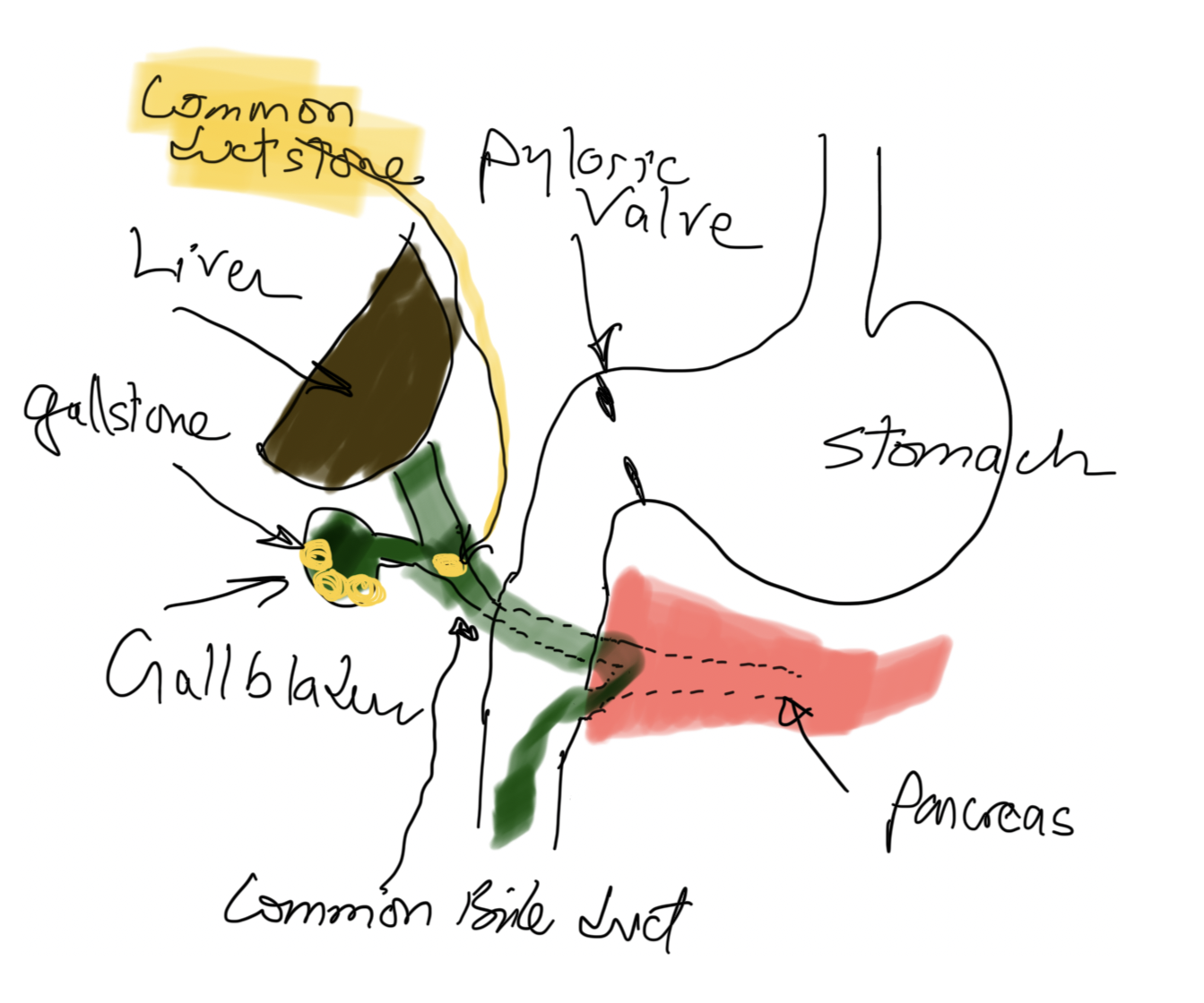
Cholangitis refers to infection and inflammation of the bile ducts because of obstruction and introduction of bacteria or other pathogens from the GI tract.
- Hydration with IV fluids,
- Broad coverage with IV antibiotics administration and
- Drainage procedure (ERCP, papillotomy, Stent placement, Cholecystectomy with common duct exploration, and T-tube placement)
These steps are almost all taking place concurrently and urgently. This is not something a patient waits and sees how it goes- it only goes one direction, from bad to worse. This is not to sound alarmist; a correct diagnosis is critical.
Risk factors for cholangitis may include gallstones that travel down the bile duct and cause jaundice, pancreatitis if passed, immunosuppression, malnutrition, and diabetes, to name a few.
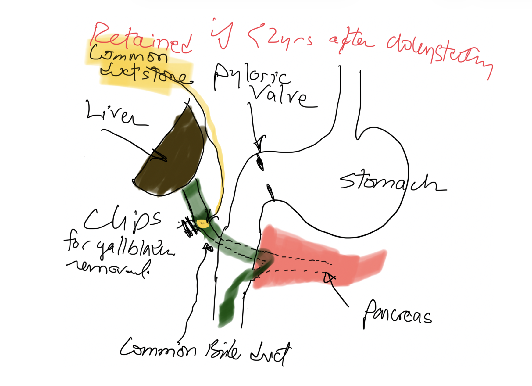
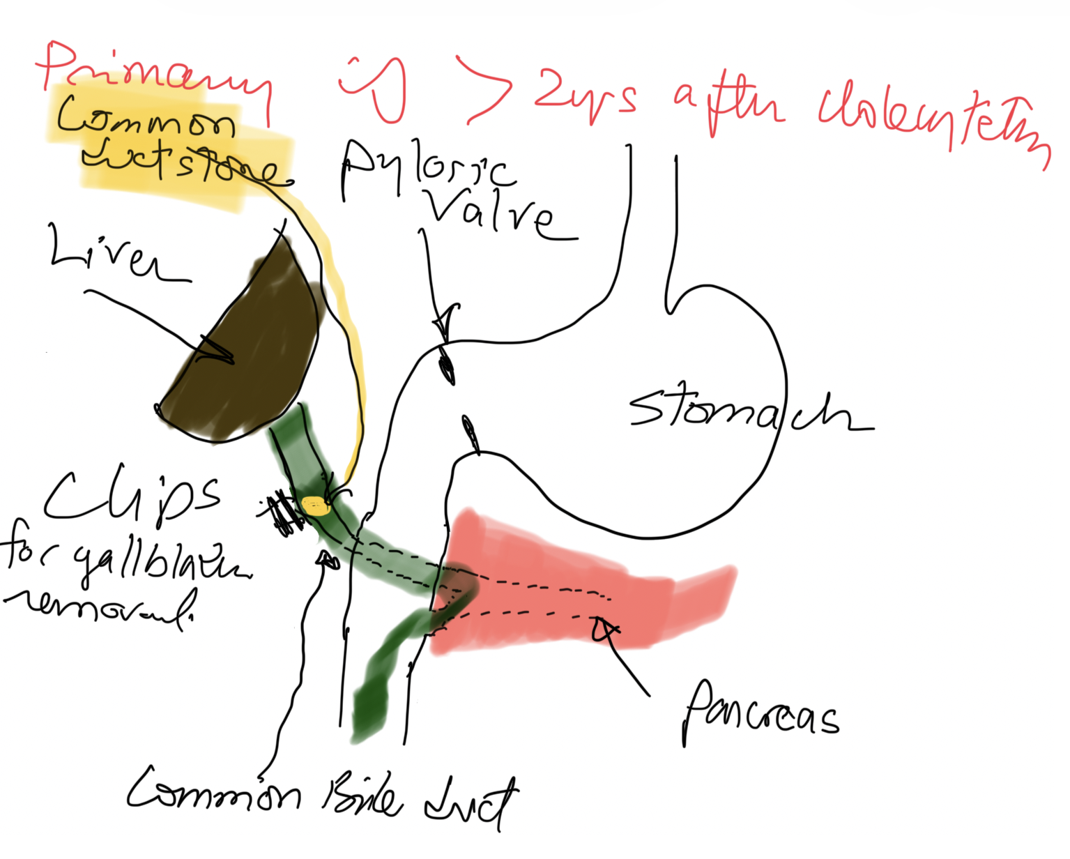
If a patient has gallstones in the bile duct less than 2 years after cholecystectomy, it is a Retained Stone(s), meaning there was a small stone that was not in the gallbladder and was not seen on US, intraoperative cholangiogram, or ERCP (if performed before surgery).
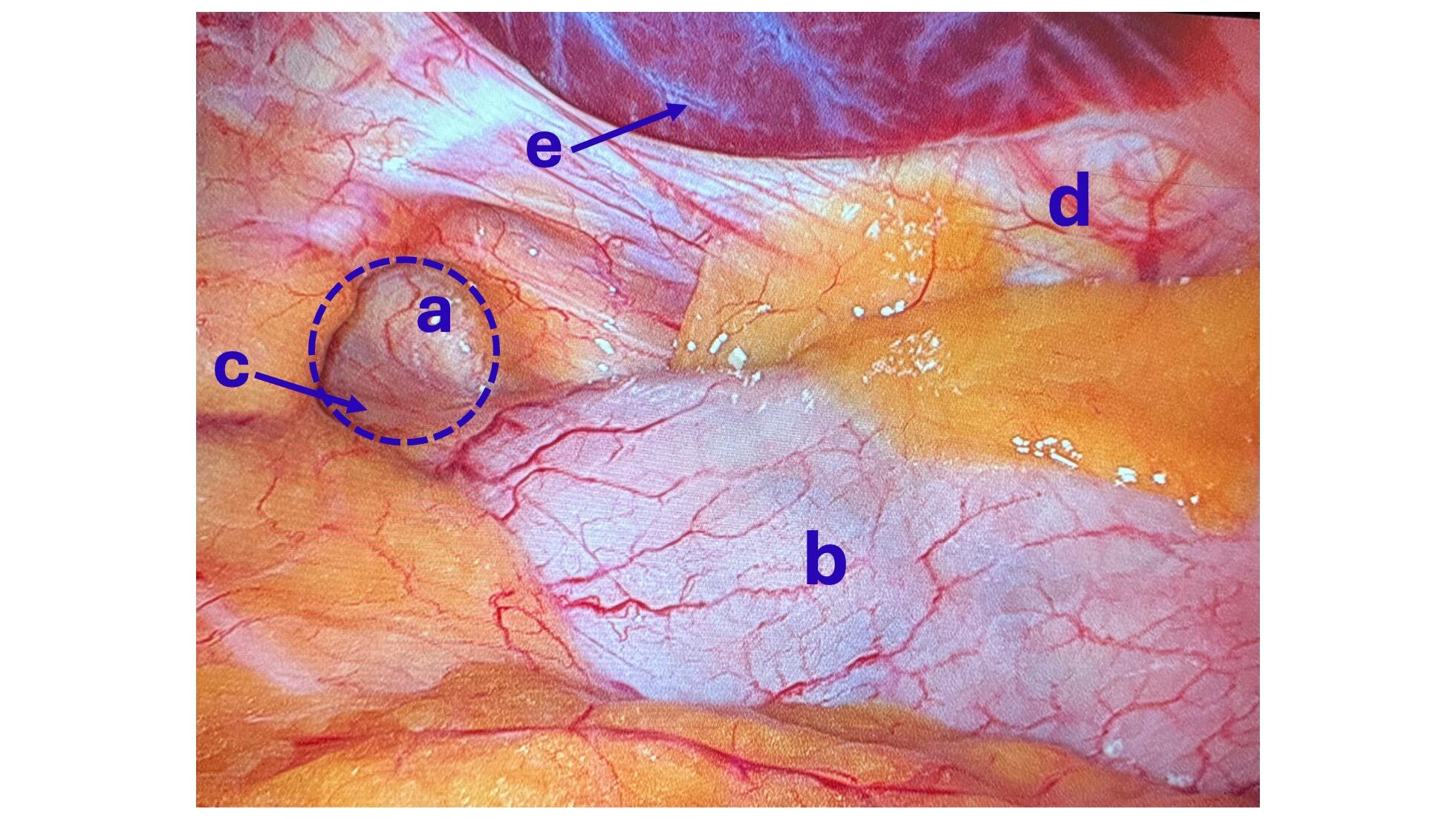
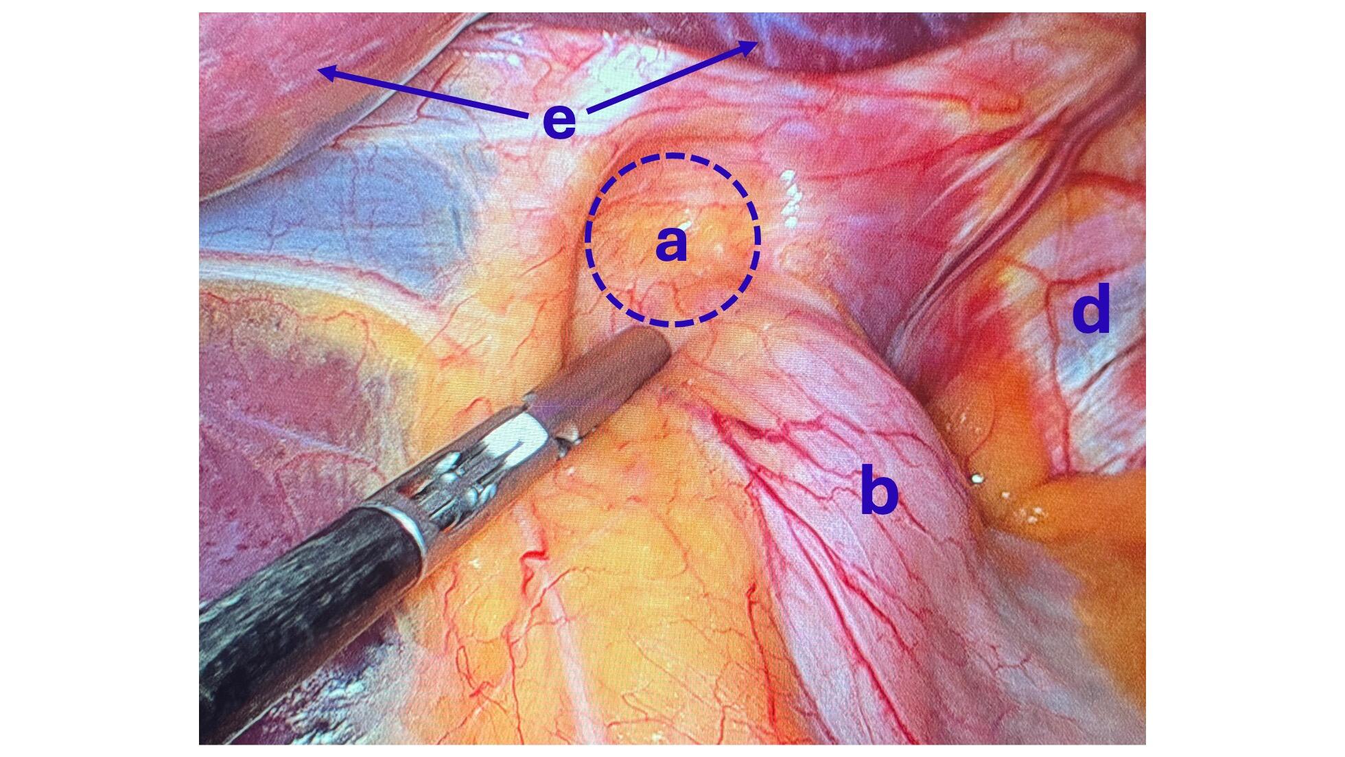
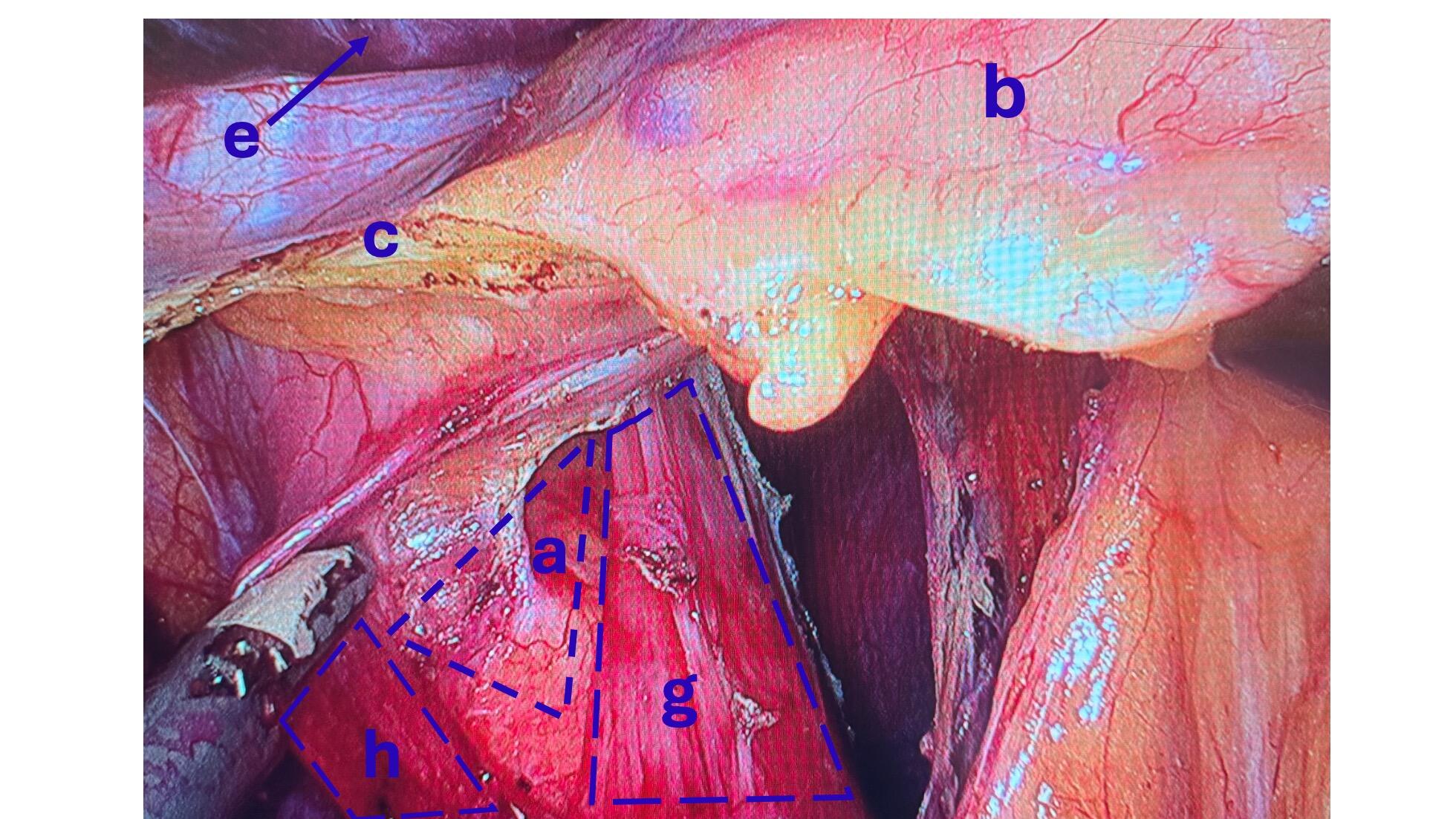
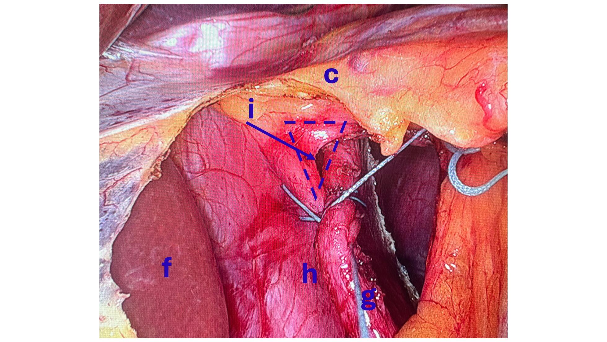
Bile Reflux Gastritis
May 19, 2026 4:13 pm
Hello, it’s best to clarify a few points:
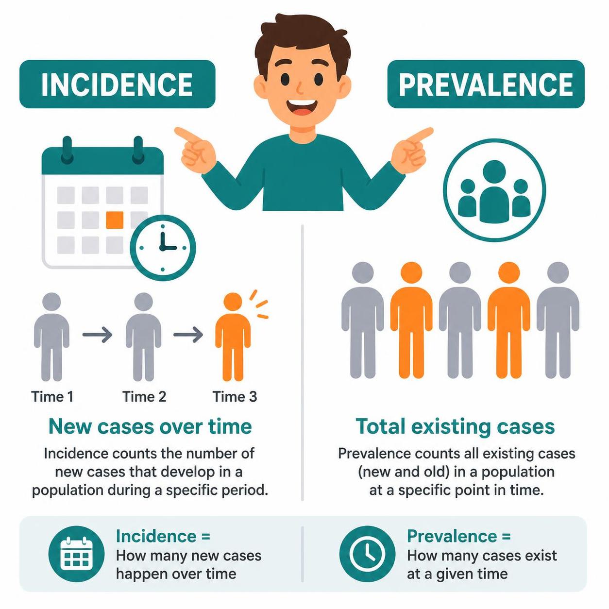
- The difference between prevalence and incidence.
- Incidence means new cases over
- Prevalence refers to existing cases at any time
- The difference between causation, coexistence, and correlation
- Causation refers to a condition causing an effect.
- Coincidence refers to two conditions present together at the same time, with neither causing the other.
- Correlation refers to two conditions that may occur together, with no causation.
- Study size refers to the research data on which the information is based. The larger the population size studied, the better the sample data and the more conclusive the results.
- Confounding factors.
Sphincter of Oddi Dysfunction
April 26, 2026 8:43 am
Description:
Sphincter of Oddi Dysfunction (SOD) is a clinical syndrome characterized by biliary/ pancreatic pain from abnormal function or obstruction of the.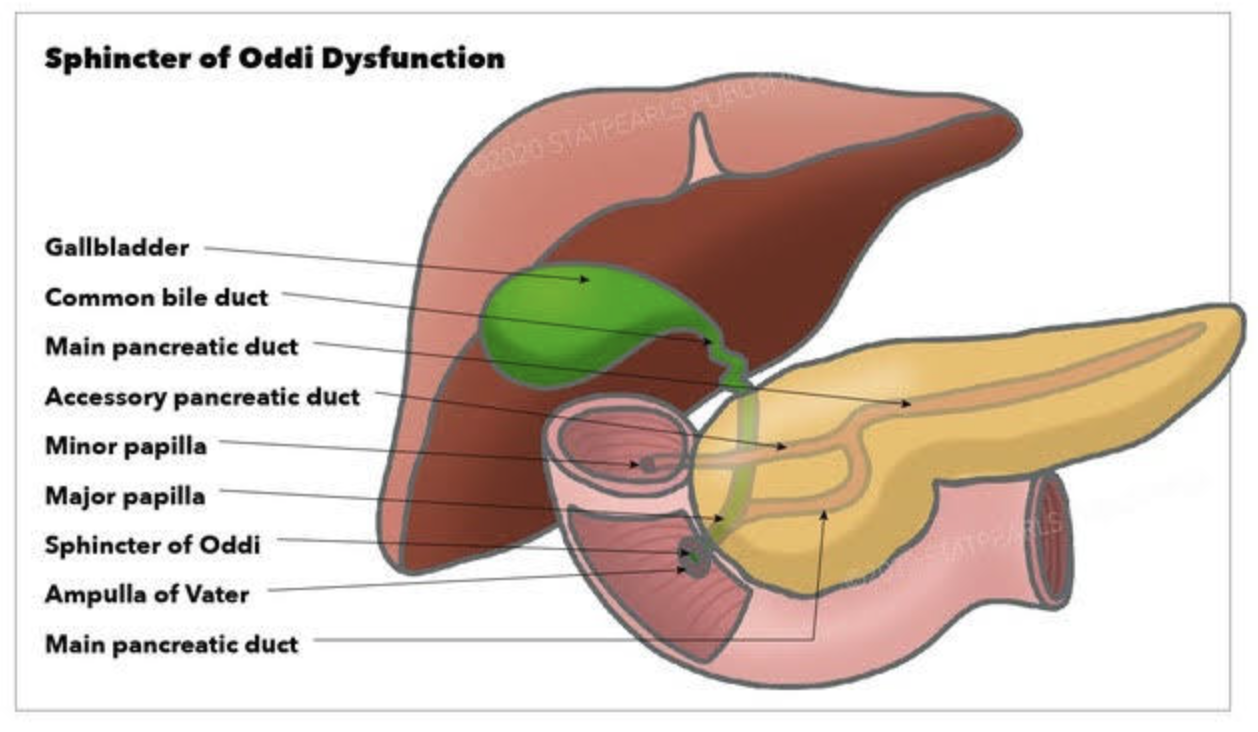
Diagnostic and classification
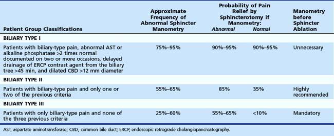
- The SOD spectrum includes biliary, pancreatic, or combined sphincter dysfunction, with symptoms driven by dyskinesia or mechanical obstruction. The term SOD encompasses both functional motility disorders (biliary or pancreatic sphincter dysfunction) and mechanical obstructions such as papillary stenosis.
- SOM has been considered the gold standard for diagnosing elevated basal sphincter pressure. It is invasive and carries complications, including pancreatitis. SOM can also not be done in those who have had gastric bypass or duodenal switch operations. Sphincterotomy outcome is not uniform. Consequently, many centers have moved toward empiric endoscopic therapy for appropriately selected patients and toward noninvasive or less invasive diagnostic approaches for others.
Treatment approaches
- Endoscopic biliary sphincterotomy (EST) is the most established nonpharmacologic treatment for biliary SOD, particularly in type I and many type II patients with objective ductal dilation and/or enzyme elevation.
- In type II SOD, outcomes after EST correlate with objective evidence of obstruction or sphincter hypertension on SOM. Empiric sphincterotomy without manometry is controversial.
- Type III SOD has emerged as predominantly functional pain rather than a mechanical obstacle. Alternative management emphasizes risk stratification, noninvasive strategies, and multidisciplinary approaches.
- Medical and non-sphincterotomy options (calcium-channel blockers, nitrates, antidepressants, and lifestyle modifications) have been discussed as potential adjuncts or alternatives.
Summary
- SOD is a multifactorial biliary and pancreatic sphincter disorder with a spectrum from mechanical obstruction to pure dyskinesia. Type I and II retain treatment relevance, particularly EST for biliary obstruction. Type II management requires clear identification of the cause.
- SOM is a diagnostic tool with significant limitations.
- Endoscopic sphincterotomy offers meaningful symptom relief in type I and select type II with objective obstruction. It carries a risk of complications, including pancreatitis; risk mitigation strategies are integral to practice. In type III SOD, sphincterotomy generally has limited benefit, necessitating a move toward noninvasive management and careful patient counseling. Patients post- gastric bypass, or duodenal switch, can not have ERCP or SOM done.
Types of HyperParathyroidism
February 20, 2026 4:05 pm
There are 4 parathyroid glands which are located behind the thyroid gland, among other functions, are the main regulators of calcium, phosphorous, and magnesium in the blood. Elevations of parathyroid hormone (hyperparathyroidism) can be: 1-Primary, 2-Secondary, 3-Tertiary

Primary hyperparathyroidism means the parathyroid glands themselves are hyperactive. This may involve only one of the four glands: a) an adenoma, a benign tumor that needs surgical removal, or b) hyperplasia, when all 4 glands are hyperactive and/or enlarged, and in some cases, most of the 4 glands need to be removed.
Secondary hyperparathyroidism means that the elevated PTH level is caused by an external regulatory stimulus, such as low calcium, which itself may be due to low vitamin D, low calcium intake, or other causes.
Tertiary hyperparathyroidism is seen only in specific renal failure and transplant patients.
Regardless of the type of hyperparathyroidism, the end result is the same. Because the parathyroid gland aims to maintain normal calcium levels, it will do everything to achieve them. This includes increasing calcium absorption from the GI tract, breaking down bone to increase the blood calcium supply, and increasing calcium reabsorption from the urine.
Distinguishing between primary and secondary is critical, as primary is more likely than not a surgical problem that needs to be addressed. Secondly, it may be responding to metabolic deficiencies (low CA, low Vitamin D) that need to be corrected and take some time.
Not all cases require surgical intervention, as labs (vitamin D, calcium, and alkaline phosphatase) and imaging studies, such as neck ultrasound, CT scan, and Sestamibi scan, provide the information needed to dictate the treatment plan. Please stay up to date with your yearly lab results to catch changes sooner rather than later.
Vitamin D level and Liver Function Test (LFT) elevation
February 08, 2026 11:21 am
After weight-loss surgery, some patients may experience a transient elevation in liver function tests that resolves over time. We have previously reported on this. IT is essential to distinguish between the Duodenal switch and the SIPS/SADI procedure, where some patients are led to believe they are identical. These procedures differ physiologically, and their weight loss and metabolic behaviours vary significantly.
Other than the stress of the weight loss, obesity, and comorbidities of obesity, there may be other anatomical post-surgical causes for elevated liver function test. This has also been discussed extensively.
A recent literature review supports the protective effects of vitamin D supplementation.
Elevated liver enzymes may be caused by many factors, including nutritional deficiencies, excessive supplementation (turmeric), medications, alcohol, adhesions causing partial bowel obstruction, and increased enterohepatic bile reabsorption . I would be very cautious about associating vitamin D supplementation with elevated liver function test results, even if the vitamin D level is in the very high normal range, regardless of the daily dose (much less frequent with injectable).
Vitamin D, as a fat-soluble vitamin, however, protects the liver and improves liver function test even in very high serum level . In rare cases, prolonged, elevated vitamin D levels may strain the liver. In Fact, the association of the vitamin D level and liver disease, including cirrhosis, leads to hepatocellular carcinoma (HCC) and dea h. Vitamin D protects the liver from HCC but cannot reduce the risk of cirrhosis.
Surgery for Sustained Weight Loss
January 07, 2026 1:31 pm
Surgery for Sustained Weight Loss
There have been numerous fad-type treatments promising easy, quick, low-risk, non-surgical treatment over the years, offered for the treatment of obesity. HCG injection, Phen-fen, Lap band, Orlistat (Xenical), Gastric balloon, GLP-1, and now endoscopic gastroplasty. The one thing they all have in common is the reported short-term outcome comparable to the long-term outcome of the surgery. Surgery has the longest history for sustained weight loss with data going back decades showing long term comorbidity resolution.
GLP-1, GIP class of medication reports the outcome 1-2-3 years compared to the 15-30 years outcome of the weight loss surgical procedures. These articles also fail to emphasize the high cost and comprehensive long-term risk with injections or pills taken daily or weekly.
Now to the latest fad: The results of the endoscopic gastroplasty are reported to be in the form of a 3-5-year interval. What these alternative low-risk treatments also have in common is the emphasis placed on short-term resolution of comorbidities and weight loss. The 5-year weight-loss rate is a dismal 11.8% (Total Body Weight Loss, %TBWL) (Lahooti et al., 2025). A 300-lb. person with gastroplasty will be down 34 lbs. in 5 years – compared to 100 lbs. following sleeve gastrectomy. Not surprisingly, the Endoscopic gastroplasty was found to have a 2.6% rate of serious adverse events (SEA) (Singh et al., 2019). Yet, the patients are under the impression that since gastroplasty is not a surgical procedure and no incisions are made, there are no serious risks.
One of the original first studies (Abu Dayyeh et al., 2022) also presented a set of data that one has to read the detailed findings to appreciate how poor the performance of the procedure is:
“Findings: Between Dec 20, 2017, and June 14, 2019, 209 participants were randomly assigned to ESG (n=85) or to control (n=124). At 52 weeks, the primary endpoint of mean percentage of EWL was 49·2% (SD 32·0) for the ESG group and 3·2% (18·6) for the control group (p<0·0001). Mean percentage of total bodyweight loss was 13·6% (8·0) for the ESG group and 0·8% (5·0) for the control group (p<0·0001), and 59 (77%) of 77 participants in the ESG group reached 25% or more of EWL at 52 weeks compared with 13 (12%) of 110 in the control group (p<0·0001). At 52 weeks, 41 (80%) of 51 participants in the ESG group had an improvement in one or more metabolic comorbidities, whereas six (12%) worsened, compared with the control group, in which 28 (45%) of 62 participants had similar improvement, whereas 31 (50%) worsened. At 104 weeks, 41 (68%) of 60 participants in the ESG group maintained 25% or more of EWL. ESG-related serious adverse events occurred in three (2%) of 131 participants, without mortality or need for intensive care or surgery.”
1-This study is about 85 patients having the endoscopic gastroplasty and 124 patient only being on diet (they compared the endoscopic gastroplasty (ESG) to diet and exercise, not sleeve gastrectomy)
2-at one year, the ESG group had lost 49.2% of the EWL (if needed to lose 100 lbs, only lost 49.2lbs)
These are examples of how patients need to educate and inform themselves beyond the headlines and ask the tough questions.
GLP-1/GIP class of medication has been promoted for everything under the sun (a little exaggeration here to make the point). Even though it may be true that many comorbidities resolve with any form of weight loss, we also know from weight-loss surgical data that many of these comorbidities return with weight gain. Which is why, in my opinion, it is deceptive to talk about the resolution of comorbidities with very little weight loss from alternative procedures such as lap band, gastric balloon, or endoscopic gastroplasty, when we know the comorbidities are resolved only when the weight loss is maintained long term.
Weight loss is maintained by Lap band adjustment (with the associated cost- and hopefully not having complications such as slipped band or continuous nausea and vomiting) or by replacing the gastric balloon every 6 months (how does that make sense), or by having the daily or weekly injection or pills of GLP-1, GIP class of medications with the risk of side-effects and the costs associated with these medications. All of this in the backdrop of near-certain weight gain when the lap band must be removed, or the gastric balloon is not reinserted, or the GLP-1 GIP medication is stopped (primarily because of the complication)
Weight-loss surgical procedures, such as Duodenal Switch, gastric sleeve, and Gastric bypass, are not risk-free. However, they have continually been shown to deliver sustained weight loss over decades.
As a surgeon with decades of experience with operating on patients with failed lap bands, endoscopic gastroplasties, GLP-1-GIP medications, and gastric balloons, one thing is common that patients say is how little they knew about their options. Patients who consider only the short-term outcome and perceive the least risk will require secondary treatment, which will deliver a more definitive result.
References
Abu Dayyeh, B. K., Bazerbachi, F., Vargas, E. J., Sharaiha, R. Z., Thompson, C. C., Thaemert, B. C., Teixeira, A. F., Chapman, C. G., Kumbhari, V., Ujiki, M. B., Ahrens, J., Day, C., Acosta, A. J., Badurdeen, D., Buttar, N. S., Clark, M. M., Eaton, L., Ghanem, O., Grothe, K., … Wilson, E. B. (2022). Endoscopic sleeve gastroplasty for treatment of class 1 and 2 obesity (MERIT): a prospective, multicentre, randomised trial. Lancet (London, England), 400(10350), 441–451. https://doi.org/10.1016/S0140-6736(22)01280-6
Lahooti, A., Westerveld, D., Johnson, K., Aneke-Nash, C., Baig, M. U., Akagbosu, C., Hanscom, M., Buckholz, A., Newberry, C., Herr, A., Schwartz, R., Yeung, M., Sampath, K., Mahadev, S., Kumar, S., Carr-Locke, D., Aronne, L., Shukla, A., & Sharaiha, R. Z. (2025). Improvement in obesity-related comorbidities 5 years after endoscopic sleeve gastroplasty: a prospective cohort study. Gastrointestinal Endoscopy, 102(1), 26–36. https://doi.org/10.1016/J.GIE.2024.12.017
Singh, S., Hourneaux de Moura, D. T., Khan, A., Bilal, M., Ryan, M. B., & Thompson, C. C. (2019). Safety and Efficacy of Endoscopic Sleeve Gastroplasty Worldwide for Treatment of Obesity: A Systematic Review and Meta-analysis. Surgery for Obesity and Related Diseases : Official Journal of the American Society for Bariatric Surgery, 16(2), 340. https://doi.org/10.1016/J.SOARD.2019.11.012
Endoscopic Gastroplasty Not the same as Lap Sleeve Gastrectomy
December 19, 2025 11:57 am
The Endoscopic Gastroplasty ESG – not to be confused with the Laparoscopic Sleeve Gastrectomy- was “endorsed” by the American Society for Metabolic and Bariatric Surgery (ASMBS) as an acceptable tool for treatment of obesity.
Patients need to understand that endoscopic gastroplasty ESG and sleeve gastrectomy LSG are not the same procedure and do not deliver the same outcome. Let’s not forget that we have seen this before with Fen Phen, Lap Band, Realize Band, Gastric balloon, GLP-1, and GIP, where low-risk, simple, and otherwise benign procedures and medications were found to be ineffective at best or to cause irreversible long-term complications at worst.
In EG, the stomach is folded, whereas in LSG, a significant portion of the stomach is removed.
The ASMBS, even in their press releas,e has highlighted:In short, while ASMBS does not consider the ESG equivalent to metabolic and bariatric surgery
(e.g., sleeve gastrectomy, gastric bypass) when it comes to expected weight loss or metabolic
impact, it does fall within a treatment continuum that includes lifestyle therapy, obesity
management medications (OMMs), and metabolic and bariatric surgery and should be available
to patients through bariatric and metabolic surgeons.
In my opinion, as supported by the published literature and the summary of the ASMBS position statement, endoscopic gastroplasty may be an appropriate procedure for some patients with the understanding that the weight loss is slower and less than that of the sleeve gastrectomy.
Surgery for Reflux
August 02, 2025 6:58 pm
Surgery for Reflux
Gastroesophageal reflux disease (GERD) is a common gastrointestinal problem in both the general population and post-weight-loss surgical patients.
The general population may have GERD symptoms with weight gain, pregnancy, hiatal hernia, Helicobacter pylori infection, gallstones, ulcers, motility issues of the esophagus or stomach, and other conditions. With proper workup, they all have their specific treatment plans. In a post-weight-loss surgical patient, some of the exact causes may be present in addition to those that may be specifically related to the type of surgery and its unique side effects and complications.
There is a subset of patients with hiatal hernia who should undergo surgical repair, but do not. There has been a series of publications from over 15 years ago that have been embraced by the gastroenterologist who frequently, incorrectly, dismisses Nissen fundoplication as an effective surgical tool for the treatment of GERD and hiatal hernia.
The treatment for GERD with a hiatal hernia, regardless of size, is Nissen Fundoplication and not lifelong use of proton pump inhibitors (omeprazole, pantoprazole) and other classes of medication. It should be noted that these medications were never approved for long-term use and can cause significant metabolic and gastrointestinal side effects.
Recent publications have shown that not only the old data that gastroenterologists frequently rely on was not reliable, but also that surgery is very safe and effective with a very low rate of short and long-term complications.
Iron Supplements
July 29, 2025 1:30 pm
Iron is critical for the production and action of red blood cells. Deficiency of iron may have many causes. These include bleeding from the GI track, inadequate absorption, inadequate intake, and menstrual losses in females.
Regardless of the underlying cause, chronic deficiency of iron will lead to iron deficiency anemia (IDA).
Normal Iron Ranges:
It’s important to have at least yearly laboratory studies to surveil for abnormalities. Normal results of iron testing may be different for men, women, and children. Iron and TIBC are measured in micrograms per deciliter (mcg/dL). Normal results for iron are:
- 65 to 175 mcg/dL for men
- 50 to 170 mcg/dL for women
- 50 to 120 mcg/dL for children
Normal results for TIBC are 250 to 450 mcg/dL for men and women.
To correct or prevent IDA, iron supplementation is recommend in some patients. Iron absorption is very inefficient. It take a long time to develop IDA and as long if not longer to correct it with oral supplementation. This is why we recommend some patient condor getting iron infusion.
For most, oral supplements are adequate. There are different formulary of iron supplement with varying degree of absorption.
Heme iron and iron bisglycinate have been shown to have much better absorption than ferrous sulfate formulary.
This is why we recommend:


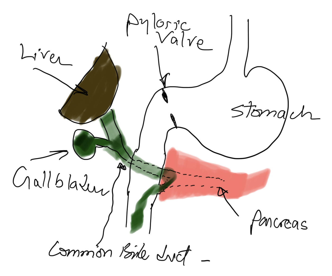
Three Different Problems: Gastritis, Gastroesophageal Reflux, and Bile reflux Gastritis
June 05, 2025 4:52 am
Gastritis is a general term used for the description of symptoms associated with several very different physical conditions and require different treatments based on their ideology . Gastritis, gastroesophageal reflux, and bile reflux gastritis
Gastritis may be caused by excess acid or bile in the stomach. Some patients may have gastroesophageal reflux due to a hiatal hernia, which needs to be treated surgically with Nissen fundoplication, regardless of the size of the hernia, contrary to what gastroenterologists recommend by prescribing antacids for an extended period. I have seen patients who have had one cm hiatal hernia and have been very symptomatic, and others who have had larger hiatal hernias and have been asymptomatic. Size should not be a determinant of whether the patient has had or will have a hernia repair or not.
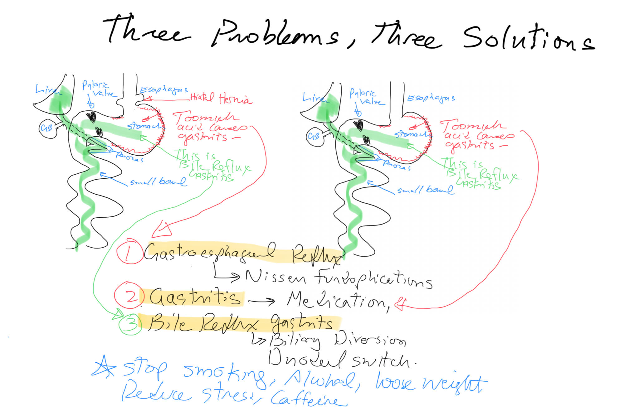
Billiary Diversion is the definitive surgical procedure for Bile Reflux Gastritis.