Category: Peristalsis
Stapled Anastomosis
December 30, 2019 11:23 am
As I was looking over old archives, I came across the following pictures that were taken years ago. These were photographs taken to demonstrate the technique for the construction of the anastomosis of the biliopancreatic channel and alimentary channel of the Duodenal Switch.
The steps of doing the stapled anastomosis of the Duodenal Switch is generally unchanged during the laparoscopic approach to the procedure.
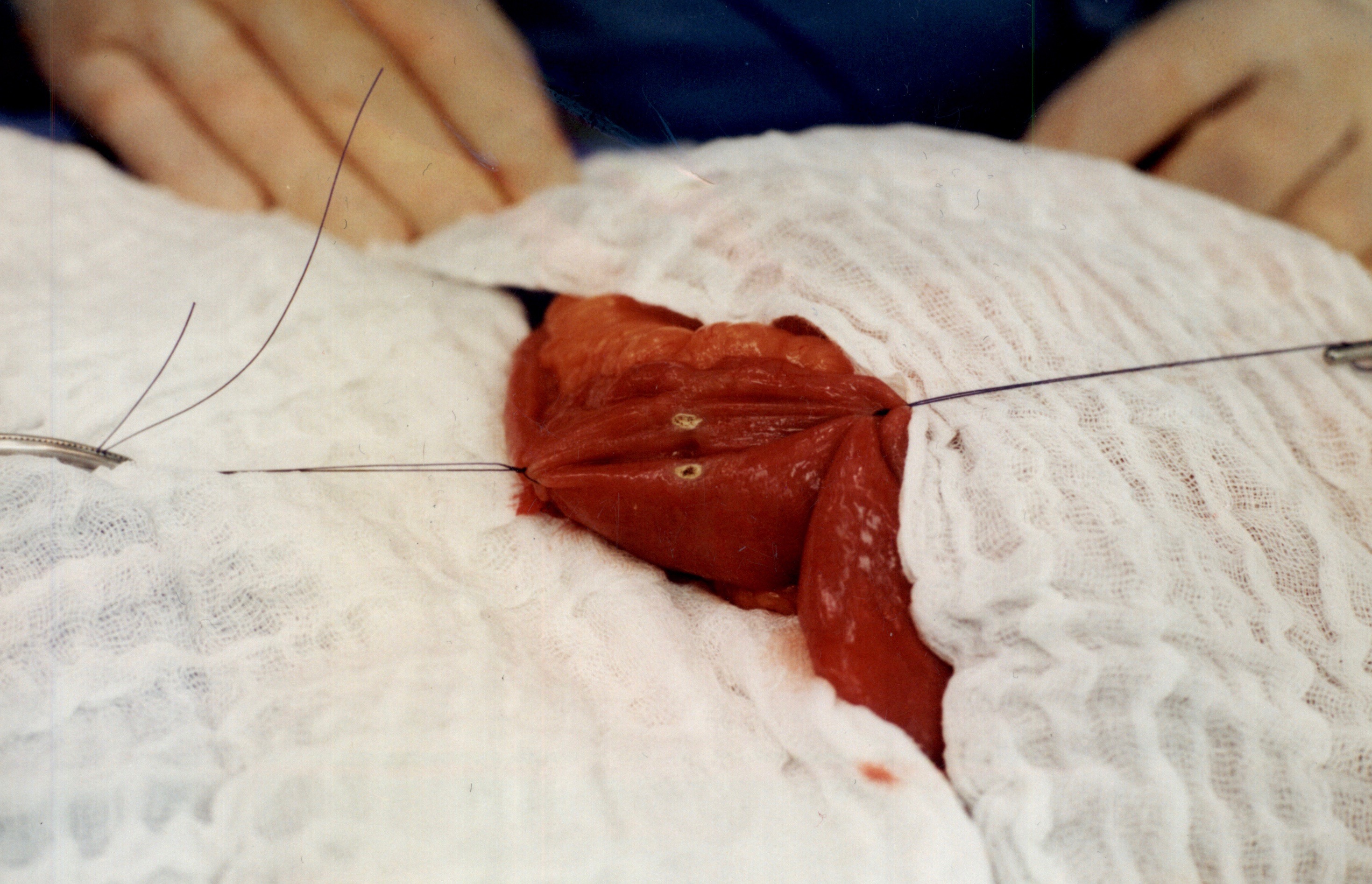
The stitches are placed to secure the bowel together. Two small openings are made in each limb of the bowel to be stapled together (the biliopancreatic limb on the bottom and the alimentary on the top of the image).
It is important to also align the bowel in the same peristalsis direction. This means that the contraction and the relaxation motion of the bowel should all point in the same direction. This should reduce the risk of complications such as intussusception.
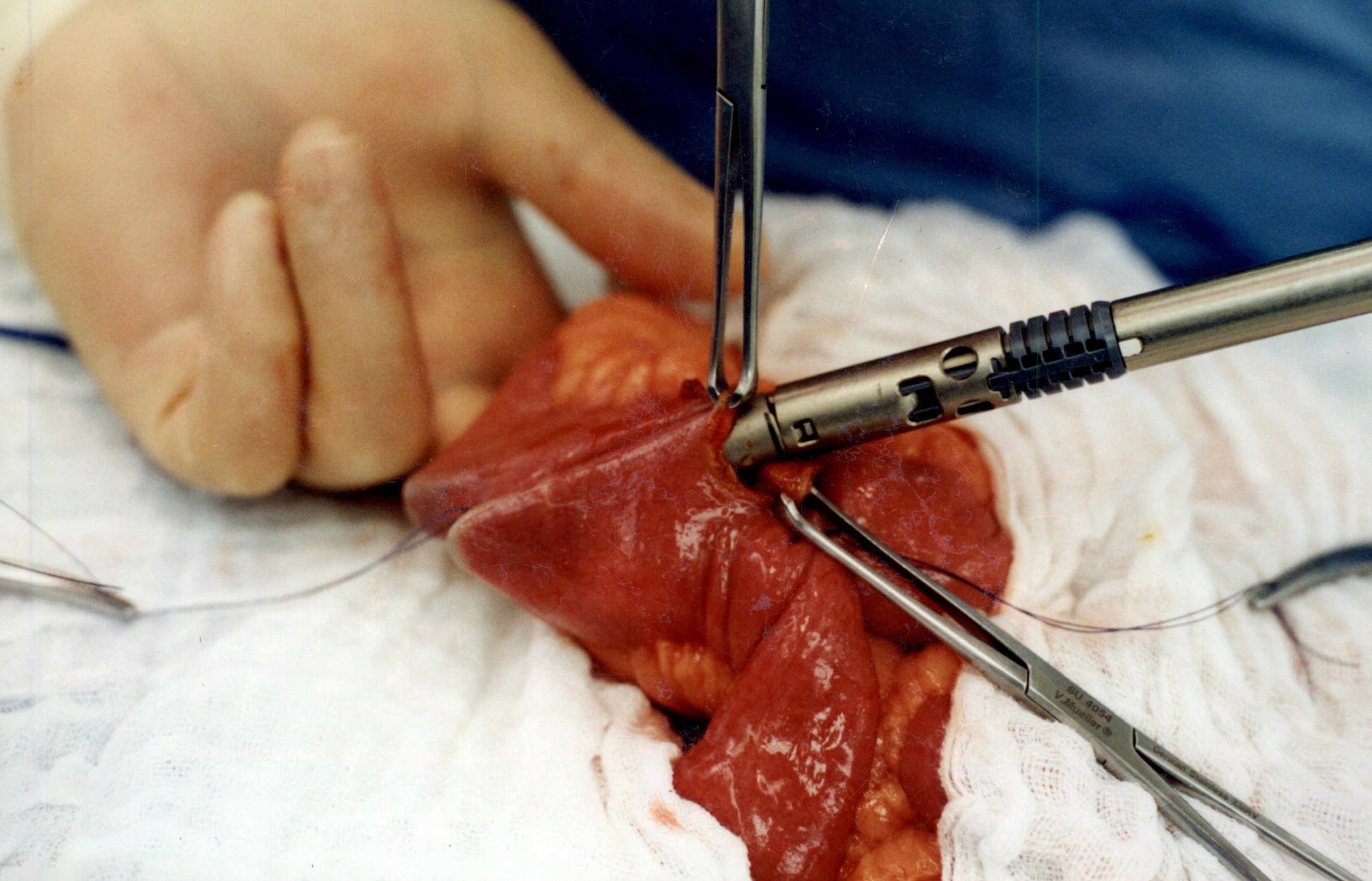
When the stapler is fired in opposite direction, a very wide anastomosis is created.
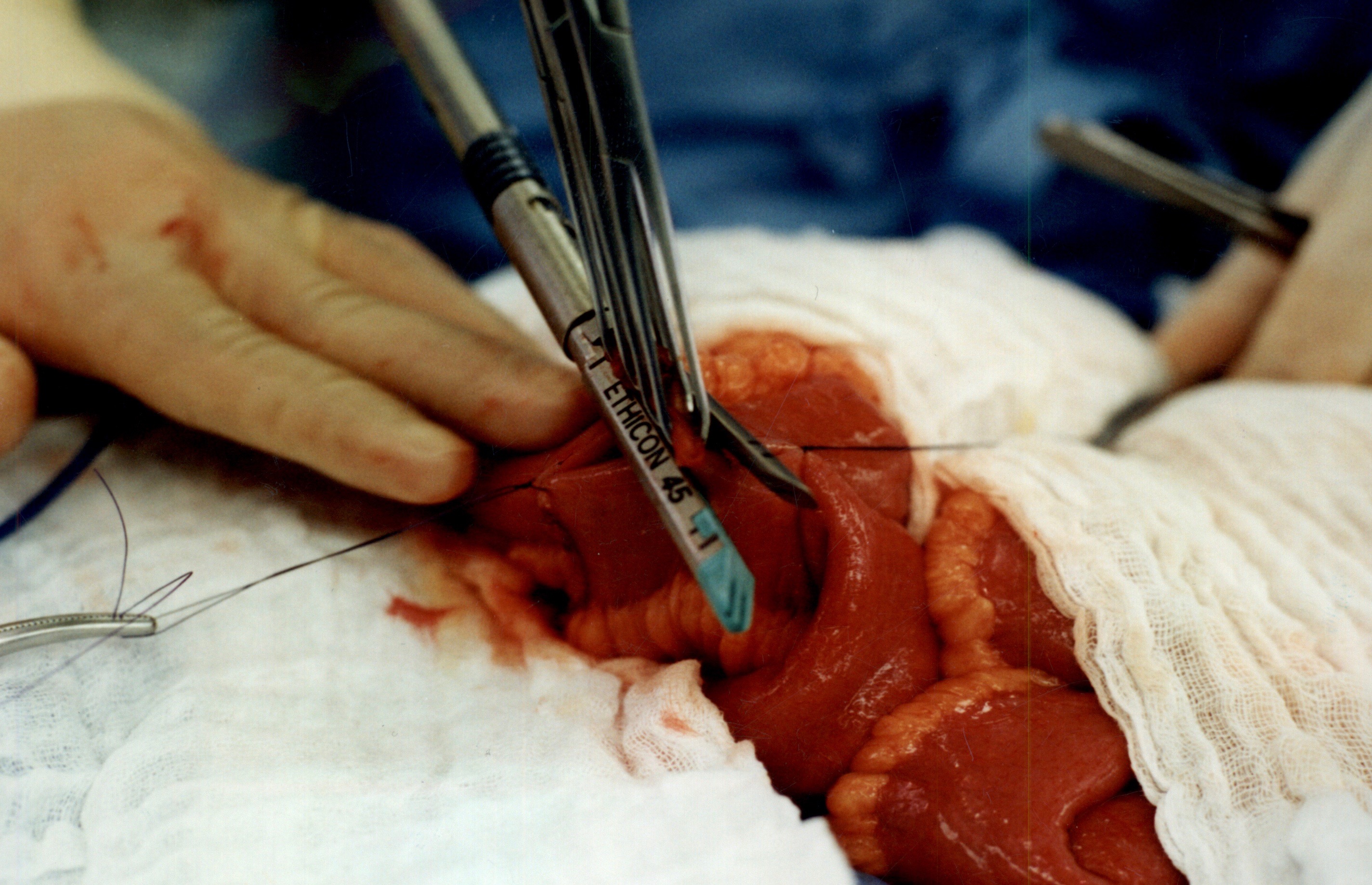
Once the anastomosis is created, then the last staple is used to close the opening that was made. This staple line is perpendicular to the direction of the anastomosis to avoid making the opening narrow.
We originally published this technique in 2003 on Obesity Surgery Journal.
Intestinal Peristalsis
June 24, 2016 6:32 pm
The following video is an example of intestinal peristalsis, the rthymic contraction and relaxation of the intestinal muscles to propel digested food through the intestinal tract. This process starts after food product is swallowed into the esophagus. It continues once the food is emptied through the pyloric valve into the small intestine. This motion allows for absorption of nutrients from the food product. Peristalsis continues throughout the small intestine and into the colon (large intestine) until defecation.
Click the following to view the Video of Intestinal Peristalsis
Peristalsis also happens within the tubes connecting the kidneys and bladder and also the tubes between the gallbladder and duodenum