
Drug Interactions Herbal Remedies
May 04, 2009 12:23 pm
Drug Interactions with Herbal remedies:
Today, one of the fastest growing supplements is the herbal remedy. Experts estimate that approximately 60-70% of the population in the United States is using some form of herbal products. What is concerning about this figure is that only a small portion of the people using herbals are communicating that fact to their health care provider. The majority of people using herbals are unaware of the potency and the interactions that herbals can have with many prescription and non-prescription medications. There are many medications that are derived from plant or herbal products such as digitalis, morphine and several anti-cancer medications. These medications easily achieve toxic levels and have very strong effects. Plant and herbs are not necessarily harmless supplements or treatments. Patients need to be aware that there is a potential for interactions with other medications and it is imperative to let your health care providers know what type of supplements or treatments you are taking over the counter.
Below is a list of a number of herbal products and their interactions with other medications both prescription and nonprescription. It is not an inclusive list and therefore anyone taking an herbal supplement or treatment should do research and always let your health care provider know what you are taking.
Patient follow up….
Follow up of patient that we have operated on takes different forms. They include review of the laboratory studies that ordered, follow up office visits, and communication with your other health care providers to name a few. None of these however are suppose to replace the routine office visits. We are very much aware of the long distances that some patient have to travel to come for their office visits. Then there are times that more time is spent waiting in our office for a relatively short visit. I can not however emphasize the importance of the a face to face office visit.
The lab results that are sent to our office are reviewed as they are received. When abnormal labs are noted appropriate measures are taken, including sending out letters that explain the abnormalities noted with recommendations that may include prescription medication. The prescriptions are also included. More frequently than not the lab results are received in our office in batches. This is why some patients receive their letters describing the deficiencies noted in their labs not all at once.
One last note regarding the follow up. We strive to be accessible at all time to patients and to their health care providers alike. We can be reached either via phone at 661-725-4847, or via email [email protected] . Every single inquiry, be from patient or another health care provider is responded to in a prompt and timely fashion.
Normal Bone Anatomy and physiology
March 11, 2009 2:42 am
Used with permission:
Susan M. Ott, MD Associate Professor, Medicine University of Washington
The bones in the skeleton are not all solid. The outside cortical bone is solid bone with only a few small canals. The insides of the bone contain trabecular
bone which is like scaffolding or a honey-comb. The spaces between the bone are filled with fluid bone marrow cells, which make the blood, and some fat
cells.

You can see the difference yourself at the grocery store meat department. Here is a photograph of a T-bone steak Below is a close-up picture of a piece of the pelvic bone. It was put into a special kind of xray machine which gives lots of details.
The photograph is used with permission from Dr. Yebin Jiang from University of California, San Francisco.
If all the bones were solid, think how heavy they would be. It would be hard to run! The next page is about the cells inside the bones.

There are three special types of cells that are found only in the bone. These cell names all start with “OSTEO” because that is the Greek word for bone.

are large cells that dissolve the bone. They come from
the bone marrow and are related to white blood cells. They are formed from two or more cells that fuse together, so the osteoclasts usually have more than one nucleus. They are found on the surface of the bone mineral next to the dissolving bone.

are the cells that form new
bone. They also come from the bone marrow and are related to structural cells. They ave only one nucleus. Osteoblasts work in teams to build bone. They produce new bone called “osteoid” which is made of one collagen and other protein. Then they control calcium and mineral deposition. They are found on the surface of the new bone.

When the team of osteoblasts has finished filling in a cavity, the cells become
flat and look like pancakes. They line the surface of the bone. These old osteoblasts are also called . They regulate passage of calcium into and out of the bone, and they respond to hormones by making special proteins that activate the osteoclasts.

are cells inside the bone. They
also come from steoblasts. Some of the osteoblasts turn into osteocytes while the new bone is being formed, and the osteocytes then get urrounded by new bone. They are not isolated, however, because they send out long branches that connect to the other osteocytes. These cells can sense pressures or cracks in the bone and help to direct where osteoclasts will dissolve the bone.

Splenda Contributes to Weight Gain
March 09, 2009 9:16 pm
This article was taken from consumeraffair.com
A new Duke University study finds that the artificial sweetener Splenda contributes to obesity, destroys beneficial intestinal bacteria and may interfere with absorption of prescription drugs.
It’s the latest in a continuing round of studies, claims and counter-claims pitting artificial sweeteners against the powerful Sugar Association, the lobbying group for the sugar industry, which financed the Duke study.
McNeil Nutritionals, which manufactures Splenda, said the study’s findings were “unsupported by the data presented” and said Splenda may be safely used “as part of a healthy diet.” The study is scheduled to be published in a forthcoming issue of The Journal of Toxicology and Environmental Health. An advance copy appears on its Web site.
A Minneapolis-based group called Citizens for Health said the Duke study demonstrates that Splenda is a health threat. The group, headed by attorney Jim Turner, has been collecting consumer reports of side effects supposedly caused by Splenda.
“The report makes it clear that the artificial sweetener Splenda and its key component sucralose pose a threat to the people who consume the product. Hundreds of consumers have complained to us about side effects from using Splenda and this study … confirms that the chemicals in the little yellow package should carry a big red warning label,” said Turner.
Turner’s group has filed a petition with the U.S. Food and Drug Administration (FDA) calling on it to review its approval of sucralose and to require a warning label on Splenda packaging cautioning that people who take medications or gave gastrointestinal problems avoid using Splenda.
“The new study makes it clear that Splenda can cause you to gain weight and lose the benefits of medications designed to improve and protect your health. The FDA should not continue to turn a blind eye to this health threat,” Turner said.
In February, a study published in Behavioral Neuroscience cites laboratory evidence that the widespread use of nocalorie sweeteners may actually make it harder for people to control their intake and body weight.
McNeil and the Sugar Association have been waging war in the courts and the public arena for years. In 2004, the association sued McNeil, claiming it had misled consumers by claiming that Splenda was “made like sugar, so it tastes like sugar.”
Splenda’s main ingredient — sucralose — is manufactured. The process involves the use of a sugar molecule but there is no sugar in the finished product.
The Duke study was conducted on rats over a 12-week period. A lead researcher, Dr. Mohamed B. Abou-Donia, said the Sugar Association had no input into the study’s findings.
In the February study, psychologists at Purdue University’s Ingestive Behavior Research Center reported that compared with rats that ate yogurt sweetened with sugar, those given yogurt sweetened with zero-calorie saccharin later consumed more calories, gained more weight, put on more body fat, and didn’t make up for it by cutting back later. Authors Susan Swithers, PhD, and Terry Davidson, PhD, theorize that by breaking the connection between a sweet sensation and high-calorie food, the use of saccharin changes the body’s ability to regulate intake. That change depends on experience.
Problems with selfregulation might explain in part why obesity has risen in parallel with the use of artificial sweeteners. It also might explain why, says
Swithers, scientific consensus on human use of artificial sweeteners is inconclusive, with various studies finding evidence of weight loss, weight gain or little effect.
Because people may have different experiences with artificial and natural sweeteners, human studies that don’t take into account prior consumption may produce a variety of outcomes.
Three different experiments explored whether saccharin changed lab animals’ ability to regulate their intake, using different assessments — the most obvious being caloric intake, weight gain, and compensating by cutting back.
The experimenters also measured changes in core body temperature, a physiological assessment. Normally when we prepare to eat, the metabolic engine revs up. However, rats that had been trained to respond using saccharin (which broke the link between sweetness and calories), relative to rats trained on glucose, showed a smaller rise in core body temperate after eating a novel, sweettasting, high-calorie meal. The authors think this blunted response both led to overeating and made it harder to burn off sweettasting calories.
“The data clearly indicate that consuming a food sweetened with no-calorie
saccharin can lead to greater body-weight gain and adiposity (fat) than would consuming the same food sweetened with a higher-calorie sugar,” the authors wrote.
The authors acknowledge that this outcome may seem counterintuitive and might not come as welcome news to human clinical researchers and health-care practitioners, who have long recommended low- or nocalorie sweeteners. What’s more, the data come from rats, not humans. However, they noted that
their findings match emerging evidence that people who drink more diet drinks are at higher risk for obesity and metabolic syndrome, a collection of medical problems such as abdominal fat, high blood pressure and insulin resistance that put people at risk for heart disease and diabetes.
Why would a sugar substitute backfire?
Swithers and Davidson wrote that sweet foods provide a “salient orosensory stimulus” that strongly predicts someone is about to take in a lot of calories.
Ingestive and digestive reflexes gear up for that intake but when false sweetness isn’t followed by lots of calories, the system gets confused.
Thus, people may eat more or expend less energy than they otherwise would.
The good news, Swithers says, is that people can still count calories to regulate intake and body weight. However, she sympathizes with the
dieter’s lament that counting calories requires more conscious effort than consuming low-calorie foods. Swithers adds that based on the lab’s
hypothesis, other artificial sweeteners such as aspartame, sucralose and acesulfame K, which also taste sweet but do not predict the delivery of calories, could have similar effects. Finally, although the results are consistent with the idea that humans would show similar effects, human
Susan E.; Davidson, Terry L. Behavioral Neuroscience. Vol 122(1), Feb 2008, 161-173.
Animals may use sweet taste to predict the caloric contents of food. Eating sweet noncaloric substances may degrade this predictive relationship, leading to positive energy balance through increased food intake and/or diminished energy expenditure. These experiments were designed to test the hypothesis that experiences that reduce the validity of sweet taste as a predictor of the caloric or nutritive consequences of eating may contribute to deficits in the regulation of energy by reducing the ability of sweet-tasting foods that contain calories to evoke physiological responses that underlie tight regulation. Adult male Sprague-Dawley rats were given differential experience with a sweet taste that either predicted increased caloric content (glucose) or did not predict increased calories (saccharin). We found that reducing the correlation between sweet taste and the caloric content of foods using artificial sweeteners in rats resulted in increased caloric intake, increased body weight, and increased adiposity, as well as diminished caloric compensation and blunted thermic responses to sweettasting diets. These results suggest that consumption of products containing artificial sweeteners may lead to increased body weight and obesity by interfering with fundamental homeostatic, physiological processes.
Confusion Regarding Duodenal Switch
March 07, 2009 12:39 pm
The duodenal switch operation includes a restrictive and malabsorptive component. This has resulted in conflicting opinions regarding the side effects of the malabsorptive component; some investigators believe that severe metabolic and nutritional complications are frequent after the operation. This likely is due to a presumed similarity of the currently performed malabsorptive operations (ie. biliopancreatic diversion and duodenal switch) to the old and now-discredited jejunoileal bypass (JIB). Unlike the jejunoileal bypass, the duodenal switch does not have a blind enteric limb.Gary J. Anthone, MD, FACS, “The Duodenal Switch Operation for Morbid Obesity”, Aug 2005, pg 820-821. [9]
Vitamin D Found To Stimulate A Protein That Inhibits The Growth Of Breast Cancer Cells
March 06, 2009 9:44 pm
Chistakos, a professor of biochemistry, has published extensively on the multiple roles of vitamin D, including inhibition of the growth of malignant cells found in breast cancer. Her current findings on the vitamin D induced protein that inhibits breast cancer growth are published in a recent issue of The Journal of Biological Chemistry. Previous research had determined that increased serum levels of vitamin D are associated with an improved diagnosis in patients with breast cancer. Prior to the current study, little was known about the factors that determine the effect of calcitrol on inhibiting breast cancer growth, she said. During the study, Christakos and coauthor Puneet Dhawan, Ph.D., examined the protein involved in the raction that can reduce the growth of vitamin D in breast cancer cells. “These results provide an important process in which the active form of vitamin D may work to reduce growth of breast cancer cells,” said Christakos. “These studies provide a basis for the design of new anticancer agents that can target the protein as a candidate for breast cancer treatment.”
Women should go for the broccoli when the relish tray comes around during holiday celebrations this season.
While it has been known for some time that eating cruciferous vegetables, such as broccoli, cauliflower, and cabbage, can help prevent breast cancer, the mechanism by which the active substances in these vegetables inhibit cell proliferation was unknown — until now.
Scientists in the UC Santa Barbara laboratories of Leslie Wilson, professor of biochemistry and pharmacology, and Mary Ann Jordan, adjunct professor in the Department of Molecular, Cellular, and Developmental Biology, have shown how the healing power of these vegetables works at the cellular level. Their research is published in this month’s journal Carcinogenesis. “Breast cancer, the second leading cause of cancer deaths in women, can be protected against by eating cruciferous vegetables such as cabbage and near relatives of cabbage such as broccoli and cauliflower,” said first author Olga Azarenko, who is a graduate student at UCSB. “These vegetables contain compounds called isothiocyanates which we believe to be responsible for the cancer-preventive and anti-carcinogenic activities in these vegetables. Broccoli and broccoli sprouts have the highest amount of the isothiocyanates. “Our paper focuses on the anti-cancer activity of one of these compounds, called sulforaphane, or SFN,” Azarenko added. “It has already been shown to reduce the incidence and rate of chemically induced mammary tumors in animals. It inhibits the growth of cultured human breast cancer cells, leading to cell death.” Azarenko made the surprising discovery that SFN inhibits the proliferation of human tumor cells by a mechanism similar to the way that the
anticancer drugs taxol and vincristine inhibit cell division during mitosis.
Mitosis is the process in which the duplicated DNA in the form of chromosomes is accurately distributed to the two daughter cells when a cell divides.
Hundreds of tiny tube-like structures, called microtubules, make up the machinery that cells use to separate the chromosomes. SFN, like the more powerful anticancer agents, interferes with microtubule functioning during mitosis in a similar manner to the more powerful anticancer drugs. However SFN is much weaker than these other plant-based drugs, and thus much less toxic. “SFN may be an effective cancer preventive agent because it inhibits the proliferation and kills precancerous cells,” said Wilson. It is also possible that it could be used as an addition to taxol and other similar drugs to increase effective killing of tumor cells without increased toxicity.
Calcium May Cut Cancer Risk
March 06, 2009 11:10 am
Calcium May Cut Cancer Risk
Higher Calcium Intake May Reduce Risk of Colorectal and Other Types of Digestive Cancers
Here’s yet another reason to bone up on calcium. It may help reduce your risk of cancer. A new study shows that older men and women who got the most calcium from food and supplements had a 16% lower risk of colorectal and other cancers of the digestive system than those who got the least calcium.
Among women, those cancer-fighting benefits were even stronger. Women who got the most calcium from food and supplements had a lower risk of all cancer and a 23% lower risk of cancers of the digestive system than those who got the least.
Calcium is already known to boost bone health, but researchers say previous studies on calcium’s effect on cancer have produced mixed results.
“Our study suggests that calcium intake is associated with a lower risk of total cancer and cancers of the digestive system, especially colorectal cancer,” write researcher Yikyung Park, ScD, of the National Cancer Institute and colleagues in the Archives of Internal Medicine.
Calcium May Lower Cancer Risk
The Institute of Medicine recommends 1,200 milligrams of calcium per day for men and women over age 50. Dietary guidelines also call for adults to eat three cups of fat-free or low-fat dairy foods, such as milk, yogurt, and cheese to meet their daily calcium needs. This does not include conditions or diseases that my limit the absorption of the Calcium such as weight loss surgical procedures.
One 8-ounce serving of skim milk or yogurt contains about 300 milligrams of calcium. Other nondairy sources of calcium include beans, broccoli, spinach, and other green, leafy vegetables.
In the study, researchers analyzed data from nearly 300,000 men and 200,000 women 50 to 71 who participated in the National Institutes of Health-AARP Diet and Health Study. Participants answered a questionnaire about the foods and supplements they ate, and cancer rates were linked through state cancer registries over seven years of follow up.
The results showed that total calcium intake was not associated with a lower risk of cancer in general in men, but women who got up to 1,300 milligrams of calcium per day had a lower risk of cancer overall.
However, total calcium intake from both food and supplements was linked to a lower risk of colorectal cancer and other cancers of the digestive system in both men and women.
Men who got the highest levels of total calcium per day through food and supplements had a 16% lower risk of these cancers than those who got the least amount per day. Women who get the most calcium per day had a 23% lower risk than those with the least reported intake per day.
Researchers conclude that “our findings suggest that calcium intake consistent with current recommendations is associated with a lower risk of total cancer in women and cancers of the digestive system, especially colorectal cancer, in both men and women.”
SOURCES: Park, Y. Archives of Internal Medicine, Feb. 23, 2008; vol 169: pp 391-401.
Opinion and Commentary: The above two articles that were taken from internet sites, reflect the continues and ever evolving nature of the science behind most of the recommendations made.
Just imagine the treatment of a patient with a disease process. Almost certainly the surgical and medical treatment that may have been recommended years ago may not be an option today.
This is why it is important for us, providers and patients alike to continue to stay vigilant with our surveillance of our health condition and adapt to any pertinent information that may have an impact on it. One of the most commonly asked question is about the type and the amount of the calcium that patients are required to take.
The issue of the types of the calcium supplement is discussed in great length in the FAQ section of our website (dssurgery.com, weightlossinla.com). The table outlines the differences between the types. The bottom line is that for most patients there may not be much of a difference as long as the patients are consistently taking them. There are cases however when we have recommended one type or another.
The most recommended dose is 2000-3000 mg per day in divided dose. Before some of you start calling or emailing us as to how come you were told to only take 1500 and not 2000, remember what I said earlier in this section that this is an ever evolving field. If you have taken 1500mg in divided doses and have had your yearly lab work with no further recommendation from us, do not make any changes unless instructed so.
The other most common concern regarding the calcium, is its relationship with kidney stones. Most common Kidney stone is Calcium Oxalate in post weight loss surgical patients. The treatments Does not include cutting back on Calcium. Calcium Oxalate search of our website will provide further detail.
Ara Keshishian Md, FACS
Fat Malabsorption
March 04, 2009 12:38 pm

Berry Compound Reduces Aging Effect
February 09, 2009 11:00 am
In a new study, aged laboratory animals that ate a diet rich in the berry and grape compound pterostilbene performed better than those in a group that did not eat the enriched diet, scientists with the Agricultural Research Service (ARS) have reported. Pterostilbene reversed measurable negative effects of aging on brain function and behavioral performance.
Neuroscientist James Joseph, psychologist Barbara Shukitt-Hale and colleagues at the Jean Mayer USDA Human Nutrition Research Center on Aging at Tufts University in Boston, Mass., collaborated on the study with chemist Agnes Rimando of the ARS Natural Products Utilization Research Laboratory in Oxford, Miss. For the two-part study, the researchers wanted to determine if pterostilbene would be effective in reversing the effects of aging on mature rats.
For the first part of the study, they tested seven stilbene compounds in cell cultures and found that pterostilbene was the most effective at preventing oxidative stress. For the second part of the study, they fed aged rats one of three diets: control, or control adjusted to include either low or high concentrations of pterostilbene. The results indicated that in aging rats, pterostilbene was effective in reversing cognitive decline and that improved working memory was linked to pterostilbene levels in the hippocampus region of the brain.
The study results are the latest in a series of ARS cell culture and animal model studies published in the last decade that shed light on relationships between various dietary components and brain function while aging. The authors noted that there are additional berry compounds showing similar
potential, which they continue to investigate in animal and cell models. The researchers followed protocols approved by the Frederick, Md.- based Association for Assessment and Accreditation of Laboratory Animal Care International and a Boston, Mass.-based Internal Animal Care Review Committee.
The study was published recently in the Journal of Agricultural and Food Chemistry.
Avocado
January 12, 2009 6:55 am
Avocado- Can I have some?
“Avocados Are Not Just a Pretty Face, They’re Good for You, Too.” According to a in the LA Times, a “functional food” is defined as one that helps maintain a healthy physique and improves the body. Now that’s a little vague, but primarily this definition is being used to single out those foods that seem to be naturally blessed with combinations of phytochemicals that may help fight certain chronic diseases such as cancer and heart disease. Now avocados have been elevated to “functional food” status. According to the director of the UCLA Center for Human Nutrition, avocados include a cholesterollowering agent and are said to act as an antioxidant. Although naturally high in fat (monounsaturated like olive oil) they can be added to diabetic and heart-healthy diets. Avocados have more potassium than a banana and contain almost 10 % of an adult’s daily requirement of iron. They are low in sodium, and provide good quantities of betacarotene, vitamins B6, C and E, folic acid and copper. Because they are a vegetable product, they have no cholesterol. The average avocado contains 112-177 calories depending on size and type. A slice of avocado on a low-fat cracker can provide extra protein as well. Bon Appetite!!
How to Calculate the amount of Calories used?
EXERCISE AND CALORIES—To determine the number of calories burned for a specific activity, enjoy the information from below and use it to help you burn calories!
| Activity | Calories per minute for a 150 pound individual |
| Aerobics | 9.2 |
| Archery | 4.4 |
| Jumping Rope | 11.0 |
| Billiards | 2.9 |
| Lying at ease | 1.5 |
| Boxing | 9.4 |
| Canoeing | 3.1 |
| Card playing | 1.7 |
| Nautilus | 6.3 |
| Free weight | 5.8 |
| Cleaning | 4.2 |
| Snow skiing | 8.1 |
| Cooking | 3.1 |
| Cycling | 4.4 |
| Squash | 14.4 |
| Swimming | 8.3 |
| Eating | 1.6 |
| Golf | 5.8 |
| Ironing | 2.2 |
| Basketball | 9.4 |
| Running (11 min. mile) | 13.1 |
| Running (9 min. mile) | 9.2 |
| Table Tennis | 4.6 |
| Fishing | 4.2 |
| Typing | 1.8 |
| Football | 9.0 |
| Volleyball | 3.4 |
| Gardening | 8.6 |
| Walking | 5.4 |
| Racquetball | 12.1 |
| CALORIES = calories per minute for a 150 pound individual
Total calories burned = (W/150) x C x M Where: W = Your weight in pounds C = Number from table above for specific activity M = Number of minutes activity performed Example: If a 180 pounds male swims for 30 minutes, the total calories burned are: Calories = (180/150) x 8.3 x 30 = 299 calories |
|
How much water is enough?
Most of us do not adequately hydrate our bodies, especially in the summertime and after we work out. This is critical for our post-op patients. To find out how much water you really need: take your weight, divide it in half and divide by 8.
Example:
A 300lb person — divided in half = 150 divided by 8 = 18 ½ glasses of water/ day.
A 150 lb person, divided in half = 75 divided by 8 = 9 glasses water/day.
Drinks containing caffeine and alcohol will only dehydrate you more.
Absorption of Minerals and Metals
January 12, 2009 6:42 am
The vast bulk of mineral absorption occurs in the small intestine. The best-studied mechanisms of absorption are clearly for calcium and iron, deficiencies of which are significant health problems throughout the world.
Minerals are clearly required for health, but most also are quite toxic when present at higher than normal concentrations. Thus, there is a physiologic challenge of supporting efficient but limited absorption. In many cases intestinal absorption is a key regulatory step in mineral homeostasis.
Calcium
The quantity of calcium absorbed in the intestine is controlled by how much calcium has been in the diet during recent periods of time. Calcium is absorbed by two distinct mechanism, and their relative magnitude of importance is set by dietary calcium “history”:.
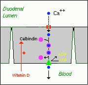
- Active, transcellular absorption occurs only in the duodenum when calcium intake has been low. This process involves import of calcium into the enterocyte, transport across the cell, and export into extracellular fluid and blood. Calcium enters the intestinal epithelial cells through voltage insensitive channels and is pumped out of the cell via a calcium- ATPase.The rate limiting step in transcellular calcium absorption is transport across the epithelial cell, which is greatly enhanced by the carrier protein calbindin, the synthesis of which is totally dependent on vitamin D.
- Passive, paracellular absorption occurs in the jejunum and ileum, and, to a much lesser extent, in the colon when dietary calcium levels have been moderate or high. In this case, ionized calcium diffuses through tight junctions into the basolateral spaces around enterocytes, and hence into blood. Such transport depends on having higher concentrations of free calcium in the intestinal lumen than in blood. Additional Calcium information here.
Phosphorus
Phosphorus is predominantly absorbed as inorganic phosphate in the upper small intestine. Phosphate is transported into the epithelial cells by contransport with sodium, and expression of this (or these) transporters is enhanced by vitamin D.
Iron
Iron homeostasis is regulated at the level of intestinal absorption, and it is important that adequate but not excessive quantities of iron be absorbed from the diet. Inadequate absorption can lead to iron-deficiency disorders such as anemia. On the other hand, excessive iron is toxic because mammals do not have a physiologic pathway for its elimination. Iron is absorbed by villus enterocytes in the proximal duodenum. Efficient absorption requires an acidic environment, and antacids or other conditions that interfere with gastric acid secretion can interfere with iron absorption.
Ferric iron (Fe+++) in the duodenal lumen is reduced to its ferrous form through the action of a brush border ferrireductase. Iron is the co-transported with a proton into the enterocyte via the divalent metal transporter DMT-1. This transporter is not specific for iron, and also transports many divalent metal ions.

Once inside the enterocyte, iron follows one of two major pathways. Which path is taken depends on a complex programming of the cell based on both dietary and systemic iron loads:
- Iron abundance states: iron within the enterocyte is trapped by incorporation into ferritin and hence, not transported into blood. When the enterocyte dies and is shed, this iron is lost.
- Iron limiting states: iron is exported out of the enterocyte via a transporter (ferroportin) located in the basolateral membrane. It then binds to the iron-carrier transferrin for transport throughout the body.
Iron in the form of heme, from ingestion of hemoglobin or myoglobin, is also readily absorbed. In this case, it appears that intact heme is take up by the small intestinal enterocyte by endocytosis. Once inside the enterocyte, iron is liberated and essentially follows the same pathway for export as absorbed inorganic iron. Some heme may be transported intact into the circulation. Additional Iron information here.
Copper
There appear to be two processes responsible for copper absorption – a rapid, low capacity system and a slower, high capacity system, which may be similar to the two processes seen with calcium absorption. Many of the molecular details of copper absorption remain to be elucidated. Inactivating mutations in the gene encoding an intracellular copper ATPase have been shown responsible for the failure of intestinal copper absorption in Menkes disease. A number of dietary factors have been shown to influence copper absorption. For example, excessive dietary intake of either zinc or olybdenum can induce secondary copper deficiency states. Additional Copper information here.
Zinc
Zinc homeostasis is largely regulated by its uptake and loss through the small intestine. Although a number of zinc transporters and binding proteins have been identified in villus epithelial cells, a detailed picture of the molecules involved in zinc absorption is not yet in hand. Intestinal excretion of zinc occurs via shedding of epithelial cells and in pancreatic and biliary secretions. A number of nutritional factors have been identified that modulate zinc absorption. Certain animal proteins in the diet enhance zinc absorption. Phytates from dietary plant material (including cereal grains, corn, rice) chelate zinc and inhibit its absorption. Subsistence on phytate-rich diets is thought responsible for a considerable fraction of human zinc deficiencies. Additional Zinc Information here